Canon Medical Systems USA
With headquarters in Tustin, Calif., Canon Medical Systems USA Inc. markets, sells, distributes and services radiology and cardiovascular systems, and coordinates clinical diagnostic imaging research for all modalities in the United States. Canon Medical Systems Corporation, an independent group company of Canon Inc., is a global leading provider of diagnostic imaging systems including CT, MRI, Ultrasound, X-ray systems and clinical laboratory systems. In business for more than 100 years, Canon Medical Systems Corporation (formerly Toshiba Medical Systems Corp.) was built to improve the quality of life for all people. It delivered on this mission with medical innovations that are “Made for Life”—made to improve the lives of patients, clinicians and administrators. Its legacy was built with pioneering medical technology, such as the world's first X-ray machines in 1932 and Japan's first magnetic resonance imaging (MRI) systems in 1983. Canon Inc. entered the healthcare business in 1940 with the development of Japan’s first indirect X-ray camera which was used for the early detection of pulmonary tuberculosis. The partnership of Toshiba Medical Systems Corporation and Canon (now Canon Medical Systems Corp.) brings together two cutting-edge technology businesses, founded with similar values of creativity, flexibility and patient-friendly healthcare solutions. Today, their combined history of research and innovation drive forward Canon Medical Systems’ vision for building a world-class healthcare enterprise. For more information: https://us.medical.canon/
Canon Medical Systems’ offering includes: computed tomography, magnetic resonance, ultrasound, X-ray, cath and EP lab
Videos
-
August 31, 2021
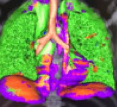
Several radiology IT vendors at 2021 Healthcare Information Management Systems Society (HIMSS) conference demonstrated computed tomography (CT) imaging advanced visualization software software to help automatically identify and quantify COVID-19 pneumonia in the lungs. These tools can help speed assessment of the lung involvement and serial tracking can be used to assess the patient's progress in the hospital and during long-COVID observation.
Examples of COVID analysis tool shown in this video include clips from booth tours at:
• Fujifilm
• Siemens Healthineers
• Canon (Vital)Canon received FDA clearance for its tool under and emergency use authorization (EUA).
Siemens said its tool was part of its lung analysis originally developed for cancer but modified and prioritized to aid in COVID assessments.
HIMSS Related Content:
Advances in CVIS and Enterprise iImaging at HIMSS 21
Photo Gallery of New Technologies at HIMSS 2021
VIDEO: Importance of Body Part Labeling in Enterprise Imaging — Interview with Alex Towbin, M.D.
VIDEO: Coordinating Followup for Radiology Incidental Findings — Interview with David Danhauer, M.D.
VIDEO: Cardiology AI Aggregates Patient Data and Enables Interactive Risk Assessments
VIDEO: Example of Epsilon Strain Imaging Deep Integration With Siemens CVIS
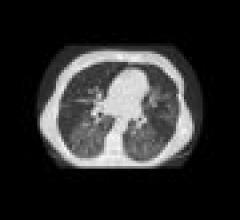
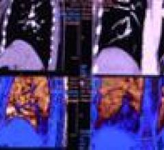
January 26, 2021This is an example of a COVID-19 (SARS-CoV-2) positive patient's lung computed tomography (CT) scan. The video scrolls through the image slices of the scan and shows the typical white, ground glass opacities (GGO) caused by COVID pneumonia. The pneumonia typically appears along the walls of each lobe of the lung, especially the chest wall and the lower portions of the lungs. This scan is from a Canon Aquilion Prime SP CT scanner and used Advanced intelligent Clear-IQ Engine (AiCE), an artificial intelligence-driven image reconstruction software to improve image quality of lower-dose scans. This was shown by Canon Medical as an exmaple of CT image quality for the virus at the 2020 Radiological Society of North American (RSNA) meeting.
Read more about this system and its launch in 2020 to address COVID, Canon Medical Launches CT Solution for Patients with Viral Infectious Diseases.
VIDEO: How to Image COVID-19 and Radiological Presentations of the Virus interview with Margarita Revzin, M.D., associate professor of radiology and biomedical imaging, Yale School of Medicine.
Find more radiology clinical images of coronavirus in this photo gallery.
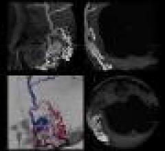
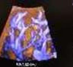
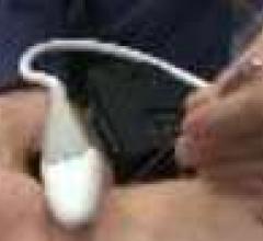
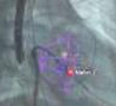
February 08, 2019This is an example of an arterial venous malformation (AVM) in the brain imaged on a Canon Alphenix Alpha angiography system. It shjows a contrast injection highlighting the vessels, which have been color coded to show the position of the veins and arteries involved in this vascular defect.
July 21, 2017DAIC and ITN Editor Dave Fornell discusses some of the most innovative new computed tomography (CT) technology and trends at the Society of Cardiovascular Computed Tomography (SCCT) 2017 meeting. Read the article "Advances in Cardiac CT Technology" and watch VIDEO: Advances in Cardiac CT Imaging.
October 05, 2016Contributing Editor Greg Freiherr offers an overview of digital radiography (DR) advances at the Association for Medical Imaging Management (AHRA) 2016 meeting. Read the article “The Coming Push for DR.” Watch a technology report sidebar video on new DR Systems technology.
December 14, 2015Video discussion of new technology and trend highlights at the Radiological Society of North America (RSNA) 2015 meeting with ITN editor Dave Fornell and ITN contributing editor Greg Freiherr.
December 11, 2015ITN/DAIC Editor Dave Fornell shows his choices for some of the most innovative new imaging technologies on the expo floor at Radiological Society of North America (RSNA) 2015 meeting.
December 11, 2015Interview with Jon Brubaker, MBA, RCVT, ultrasound technology analyst, MD Buyline, explains the trends and new technology he saw at the Radiological Society of North America (RSNA) 2015 meeting.
July 18, 2014The Aplio 500 CV is the system of choice for all premium 2-D cardiac exams. Featuring Toshiba's 2-D Wall Motion Tracking technology, the system provides stunning visualization and quantitative analysis of myocardial wall motion with unrivaled accuracy and reproducibility. With on-board cardiac quantification measurements in all directions (radial, circumferential, 2D rotation and longitudinal), the Aplio500 CV system is designed to get the most comprehensive information anytime and anywhere in the hospital, right at the patient's bedside. Additional cardiac-specific technologies include Tissue Enhancement, Advanced Dynamic Flow, Lateral Gain Controls, Tissue Doppler, Stress Echo, Flex-M Mode and Auto IMT. The system is easy to use, with superior ergonomics and a smaller footprint, making it easier to maneuver for greater patient access and improved workflow. For more information: http://medical.toshiba.com/products/ul/cardiovascular/index.php
July 03, 2014DAIC Editor Dave Fornell highlights his choices for some of the most innovative new technology at the American Society of Echocardiography (ASE) 2014 meeting.
June 20, 2014DAIC Editor Dave Fornell shares his choices for the most innovative new technologies in nuclear imaging that were on display at the 2014 Society of Nuclear Medicine and Molecular Imaging (SNMMI) annual meeting.
July 15, 2013DAIC Editor Dave Fornell highlights some of the biggest trends and most innovative technology discussed during the American Society of Echocardiology (ASE) 2013 annual meeting.
RELATED CONTENT
 News
NewsJuly 27, 2012 — The Journal of Ultrasound in Medicine has published a pilot study conducted at Thomas Jefferson University in Philadelphia, with funding provided by Toshiba, that explores the effectiveness of Toshiba America Medical Systems Inc.’s MicroPure image processing technique in visualizing breast microcalcifications compared with grayscale ultrasound imaging.
 July 27, 2012
 Feature | Dave Fornell
There are currently three major trends in cardiovascular computed tomography (CT) technology — lowering radiation dose, development of myocardial perfusion imaging and the use of CT datasets for procedural planning, especially for transcatheter aortic valve replacement (TAVR).
July 17, 2012
 News
NewsJuly 10, 2012 — In CT (computed tomography) imaging, healthcare facilities strive to limit patient radiation dose while maintaining high image quality for accurate diagnoses. St. Elizabeth Healthcare, in Edgewood, Ky., is the first facility to be upgraded with Toshiba’s advanced dose reduction software, AIDR 3D, on its Aquilion ONE CT system.
July 10, 2012
 Feature | Dave Fornell
With last year’s U.S. Food and Drug Administration approval of transcatheter aortic valve replacement (TAVR) and increasingly complex cath lab and hybrid operating room (OR) interventions, ultrasound is playing an increasing role in procedural guidance. This is especially true for use of 3-D/4-D transesophageal echo (TEE), which enables the more precise surgical-view visualization required for structural heart procedures, such as atrial septal defect (ASD) occluder and MitraClip deployments. These repairs would otherwise be extremely difficult to accomplish using fluoroscopy alone.
July 06, 2012
 News
NewsJune 29, 2012 — More than 61 companies will display their latest products and services at the American Society of Echocardiography (ASE) Annual Scientific Sessions, June 30-July 3 in National Harbor, Md. Highlights from the show floor, as reported by the vendors, will include:
June 29, 2012
 News
NewsJune 25, 2012 — KLAS released the 2012 Best in KLAS Awards: Medical Equipment and Infrastructure report, which details what end-users think about imaging systems from specific vendors. The report recognizes leaders in individual medical equipment market segments as Best in KLAS, including digital X-ray, CT, MRI (1.5T and 3.0T), ultrasound and mammography.
June 25, 2012
 News
NewsJune 22, 2012 — Toshiba America Medical Systems Inc. has partnered with United Medical Instruments (UMI) to sell Toshiba’s ultrasound systems to the private physician office and clinic market in the United States.
June 22, 2012
 News
NewsJune 18, 2012 — Toshiba America Medical Systems, Inc. will showcase the Aplio 500, Aplio 300 and Viamo ultrasound systems at the Society for Vascular Ultrasound (SVU) annual conference, June 7 to June 9, 2012, in National Harbor, Md. All systems combine advanced visualization capabilities with ergonomics and flexibility to improve departmental efficiency.
June 18, 2012
 Technology
June 15, 2012 — To enable hospitals to keep pace with the latest technological advances and offer better patient care and improved workflow while containing costs, Toshiba America Medical Systems, Inc. announces the availability of VeloCT.
June 15, 2012
 Feature | Dave Fornell
As catheter-based, minimally invasive procedures expand rapidly beyond treatment of the coronary arteries into all areas of the human anatomy, angiography X-ray imaging systems have moved beyond their original purpose of simple vascular imaging. Today, in addition to interventional cardiology, cath labs are frequently shared by numerous specialists, including interventional radiology, interventional oncology, electrophysiology (EP), vascular surgeons and neurology.
June 01, 2012
© Copyright Wainscot Media. All Rights Reserved. Subscribe Now