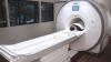
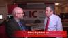
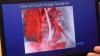
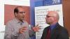
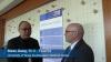
Mahadevappa Mahesh, Ph.D., chief of medical physicist and professor of radiology and medical physics, Johns Hopkins University, Baltimore, treasurer of the American Association of Physicists in Medicine (AAPM),a board member of the American College of Radiology (ACR), presented a late-breaking study on how medical imaging radiation dose has started to drop over the past decade. He is the co-chair of the National Council on Radiation Protection and Measures Report (NCRP), and presented the most recent NCRP data analysis at the 2019 Radiological Society of North America (RSNA) meeting.
The new NCRP 184 report covers the period between 2006 and 2016, the period of the most current CMS data. It shows a decrease of about 20 percent in the radiation dose the U.S. population receives from medical imaging, compared to the NCRP 160 that covered the period of up to 2006.
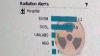
Key findings of the study include:
• CT dose dropped about 6 percent, despite a 20 percent increase CT scans since 2006;
• Drop of more than 50 percent for nuclear imaging scans, mainly due to fewer procedures being performed;
• A 15-20 percent decrease across X-ray imaging modalities.
Mahesh says this shows the impact of using "as low as reasonably achievable" (ALARA) principals, new dose guidelines outlined jointly by numerous medical societies, and dose reduction initiatives like Image Wisely, Image Gently, and the American College of Radiology (ACR) Dose Index Registry.
He said there was growing concern a decade ago when the last council report was published, which showed a steep increase in radiation dose. This was mainly due to a rapid increase in the use of computed tomography (CT) and other types of X-ray based and nuclear radiotracer medical imaging. This prompted the ACR to create the Image Wisely program and push for the use of more thoughtful imaging doses based on patient size, using the "as low as reasonably achievable” (ALARA) principle. While CT dose was lowered, he said the biggest decline over all was in nuclear imaging.
Related Medical Imaging Radiation Dose Resources:
VIDEO: Radiation Dose Monitoring in Medical Imaging — an interview with Mahadevappa Mahesh, Ph.D.
The Basics of Radiation Dose Monitoring in Medical Imaging
How to Understand and Communicate Radiation Risk — Image Wisely
Radiation in Medicine: Medical Imaging Procedures
FDA White Paper: Initiative to Reduce Unnecessary Radiation Exposure from Medical Imaging
Radiation Dose in X-Ray and CT Exams
Radiation Dose from Medical Imaging: A Primer for Emergency Physicians
Radiation risk from medical imaging
FDA: Medical X-ray Imaging
Find RSNA news and other videos
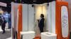
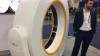
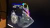
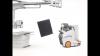
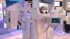
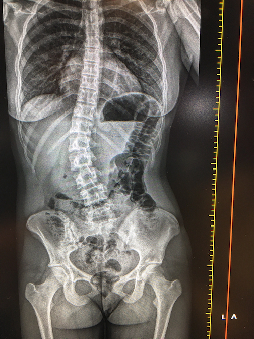 This is a demo of the EOS orthopedic X-ray imaging system at the recent 2019 Radiological Society of North America (RSNA) meeting. The system allows for weight bearing exams. EOS Imaging System the first technology capable of providing full-body, 2-D and 3-D images of pediatric patients in a standing position at a low dose of radiation. The vendor said the scans are much lower dose than computed tomography (CT).