
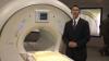
Eliot Siegel, M.D., professor and vice chairman of radiology at the University of Maryland, School of Medicine, department of radiology, discusses the big data trend, and what it means to personalized medicine as well as precision medicine.
itnTV
VIDEO: One on One with Reed A. Omary, MD, MS, Vanderbilt University Medical Center
Radiology Business | July 30, 2024
Find actionable insights to achieve sustainability and savings in radiology in this newest of ITN’s “One on One” video series with Reed A. Omary, MD, MS, Vanderbilt University Medical Center (Nashville, TN). Tune in to "Promoting the Planet's Health: Sustainability in Radiology," to hear from a recognized leader about impactful, cost-saving initiatives radiologists, associations, healthcare systems and vendors can take, and why action is imperative.
Omary, the Carol D. and Henry P. Pendergrass Professor in the VUMC Department of Radiology, is a distinguished radiologist whose commitment to driving healthcare sustainability initiatives has gained both attention and momentum. After serving as Chair of the Department of Radiology and Radiology Sciences from 2012-2023, in June, 2023, Omary stepped away from his role as Chair to pursue a sabbatical focused on climate change and sustainable healthcare. He is author of The Green Leap, a blog about making healthcare sustainable, and founder of the Greenwell Project, a sustainable healthcare non-profit. He has presented a Plenary Lecture at the Radiological Society of North America (RSNA) Annual Scientific Sessions and American College of Radiology (ACR) meetings on the topic, and continues to connect with healthcare systems, vendors and colleagues to advance the issue.
Related content:
PHILIPS MARKS MILESTONE DURING HELIUM-FREE MRI INSTALLATION IN PUERTO RICO
RSNA 2022 PLENARY SPEAKER OMARY URGES RADIOLOGISTS TO SUPPORT PATIENTS, COMMUNITIES AND THE PLANET
RSNA 2022 PANEL DISCUSSIONS FORECAST RADIOLOGY IN 2027, AND HIGHLIGHT WHY MENTORS MATTER
Radiology Imaging
Information Technology | May 30, 2014

Raym Geis, M.D., SIIM chair, spoke about how SIIM is changing its focus away from radiology and more toward informatics of all medical images. Now that every modality is starting to de-silo images, what are the next steps toward standardization?
Information Technology | May 28, 2014
Donald Dennison, SIIM Board of Directors and Hackathon organizer, discusses this inaugural event at the Society for Imaging Informatics in Medicine (SIIM) 2014 annual meeting in Long Beach, Calif.
Breast Imaging | March 27, 2014

Gary Levine, M.D., president of the National Consortium of Breast Centers, predicts a future of personalized breast screening and addresses related challenges that will need to be addressed for successful implementation.
Radiation Dose Management | March 10, 2014
GE Healthcare is dedicated to helping healthcare organizations build a roadmap for a comprehensive radiation dose management strategy. Learn about current trends in dose management and how GE Healthcare's DoseWatch solution can help you.
Computed Tomography (CT) | January 17, 2014
The BodyTom is the latest development in NeuroLogica's portable computed tomography imaging line. It is a portable, full body, 32 slice CT that boasts an impressive 85 cm gantry and 60 cm field of view. The battery powered BodyTom can be transported from room to room and is compatible with PACS, surgical navigation, electronic medical records, and planning systems. Its unique capabilities provide high quality CT images wherever needed, including the clinic, ICU, OR and emergency/trauma department. The combination of rapid scan time, flexible settings and immediate image viewing makes the BodyTom a valuable tool to any facility needing versatile real time CT imaging.
Radiology Imaging | January 14, 2014
David Widmann, president and COO of Konica Minolta Medical Imaging, discusses the company's commitment to primary imaging solutions within the diagnostic imaging market with ITN. He also shares insight into the company's entrance into the ultrasound market with the launch of the Sonimage P3 hand-held ultrasound system and acquisition of Panasonic Healthcare's ultrasonic diagnostic equipment business, as well as the recently announced global distribution agreement with GE Healthcare for Konica Minolta's AeroDR cassette-size digital X-ray imaging retrofit.
Radiology Imaging | January 14, 2014

IBA Dosimetry is excited to launch its new Digital Imaging Analysis Software IQ Analyzer Primus at the RSNA 2013. Image Quality assurance can be achieved in three simple steps: Select-Analyze-Report. The IQ Analyzer Primus can perform, fast, quantitative and reproducible constancy measurement on multiple imaging modalities, including CR, DR and RF systems. IQ Analyzer Primus provides an efficient selection and comparison of clinical phantom images from your local network and PACS system. The software includes a DICOM header tool for fast and easy selection of images. Evaluations of six individual IQ parameters is performed automatically in less than one minute. The parameters include positioning, signal to noise ratio, uniformity, dynamic scale, high contrast resolution and geometrical distortion. Users definable IQ tolerances in both absolute and percentage values, allow for simple red, yellow, and green color coded pass / fail criteria. IQ Analyzer Primus reports are available in both PDF and MS Excel formats.
Radiology Imaging | December 13, 2013

ITN Editor Dave Fornell and contributing editor Greg Freiherr discuss some of the most interesting new developments on the show floor at the Radiological Society of North America (RSNA) 2013 annual meeting.
Radiology Imaging | December 11, 2013

Greg Freiherr speaks from the show floor on day four of the 2013 annual meeting of the Radiological Society of North America (RSNA).
Radiology Imaging | December 03, 2013

Greg Freiherr speaks from the show floor on day three of the 2013 annual meeting of the Radiological Society of North America (RSNA).
Radiology Imaging | December 20, 2012
At RSNA 2012, Hitachi featured its Echelon Oval 1.5T MRI system, which features the widest bore on the market at 74 cm, a wide table and the ability to perform non-contrast MR angiography exams. Hitachi also highlighted new features for its Scenaria CT system, which is upgradeable to a 128-slice system, offers new, faster iterative reconstruction software and cardiac imaging packages.
Cardiac Imaging | December 18, 2012

ITN and DAIC Editor Dave Fornell highlights the latest advancements that will impact cardiovascular imaging from the 2012 Radiological Society of North America (RSNA) meeting. RSNA is the largest medical imaging show in the world and most advancements are shown here first.
Radiology Imaging | December 14, 2012

ITN Editor Dave Fornell highlights his choices for the most innovative radiology technologies and trends at Radiological Society of North America (RSNA), 2012. Choices include the first wireless ultrasound transducer, noiseless MRI, a 640-slice CT scanner and a printer than creates sculptures from 3-D datasets.
Find more current news and video from RSNA
Quality Assurance (QA) | December 07, 2012

At RSNA 2012, IBA showcased several quality assurance (QA) solutions for radiology. The MagicMax is an all-in-one QA system for all X-ray systems, including digital radiography (DR), CT and mammography. The Primus L phantom is an all-in-one QA device for all digital X-ray imaging systems. IBA also offers the LXcan to QA diagnostic-quality black and white flat panel monitors, and the LX Chroma to QA color displays.
Computed Tomography (CT) | December 07, 2012
Neurologica demonstrated its BodyTom portable whole-body CT scanner during RSNA 2012. The system is the first mobile scanner that can be moved on casters from room to room and is battery powered. The system has an 85 cm gantry and a 60 cm field of view. Unlike traditional CT scanners, the gantry moves over the patient, rather than the patient table being moved through the gantry. This facilitates use in the operating room, where it is not easy to move a patient who may be connected to several devices.
Radiology Imaging | December 07, 2012

Imaging Technology News experts discuss the trends and latest technology they saw on the show floor and in sessions at Radiological Society of North America (RSNA), 2012. Their discussions include some of the most innovative new devices and software to solve issues facing radiology today.
Radiology Imaging | November 12, 2012

Imaging Technology News talks to Mark Watson, executive director of the Radiological Society of North America (RSNA), and Steve Drew, RSNA's executive director for scientific assembly and informatics, about the upcoming RSNA 2012 event, as well as what's ahead for radiologists in 2013.
Digital Radiography (DR) | September 19, 2012
Carestream is changing the DR game and putting you in control of the move to digital.
Digital Radiography (DR) | September 19, 2012

The Carestream DRX-Revolution is a mobile X-ray system on wheels powered by a wireless DRX detector.
Digital Radiography (DR) | September 19, 2012
Carestream's DRX - transportable / field portable X-ray unit is designed and tested for the rigorous conditions of military, disaster and remote locations.
Computed Tomography (CT) | August 28, 2012

Web Stayman, Ph.D., Johns Hopkins University, presents an overview of research he presented at the 2012 American Association of Physicists in Medicine (AAPM) annual meeting in Charlotte, N.C. It involves an iterative technique for computed tomography (CT) to better contend with implants to improve image-guided surgery or interventions. The technique takes knowledge about the components and integrates it into the reconstruction to eliminate artifacts.
Advanced Visualization | February 13, 2012
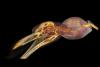
The Chicago Zoological Society's (CZS) Brookfield Zoo is the first North American zoo to use 3-D advanced visualization imaging technology. This video shows a video fly-through of reconstructed 3-D computed tomography (CT) images of an aardvark, Humboldt penguin and African crested porcupine. The zoo is using Web-based software from Vizua to create animal CT scan advanced visualization reconstructions. Read the related article.
Cardiac Imaging | December 21, 2011

ITN Editor Dave Fornell takes a tour off some of the most innovative new cardiac imaging technology advances at the Radiological Society of North America (RSNA) 2011 annual meeting.
AAPM | August 15, 2011

Tony Seibert, Ph.D., president of the American Association of Physicists in Medicine (AAPM), explained the key initiatives of the group during its 2011 annual meeting. These include:
• AAPM is working with its members to reduce patient radiation dose across radiology modalities.
• It is developing physics-based Web training modules for diagnostic radiology residents.
• Members are working to create residency programs for both radiation therapy and diagnostic radiologists.
• AAPM is also working with several states to create license certification programs to ensure who is a qualified medical physicist.
For more information: www.aapm.org
AAPM | August 15, 2011

The American Association of Physicists in Medicine (AAPM) is taking steps to help guide the future of its specialty, said AAPM President Tony Seibert, Ph.D. During the group's 2011 meeting, Seibert explained there is a shift in healthcare priorities from research to a more clinical emphasis. AAPM is encouraging younger members to get involved and keep research as an important part of medical physics, so advances can be made to eventually improve patient care.
Imaging is expanding its role in radiotherapy systems, which will require additional medical physics in that area, he said. In addition, AAPM is working with both government agencies and industry in efforts to push forward new protocols, and technology.
For more information: www.aapm.org
Radiology Business | March 22, 2011

Paul Chang, M.D., professor of radiology, vice chair of radiology informatics and medical director for enterprise imaging, University of Chicago, is a lead investigator on a closed-loop imaging research study that looks at all stages of imaging to optimize the imaging system at a hospital. The goal of the Philips-sponsored trial is to reduce errors and improving quality care and outcomes. He said it is important to optimize all stages of the imaging process. Chang explains the process they used for reviewing efficiencies and inefficiencies in the radiology department.
Watch another interview with Chang in the 2019 VIDEO: How Hospitals Should Prepare for Artificial Intelligence Implementation.
AAPM | March 22, 2011
American Association of Physicists in Medicine (AAPM) President Mike Herman, Ph.D., radiation oncology medical physicist, Mayo Clinic, explains the role of the society and its goal to improve patient care. Activities include sharing the latest scientific research, developing best practices, education, setting guidelines for certification and the roles of various staff under mediacl physicists, and how physicists can better serve their hospitals. The main focus in sessions at the AAPM annual meeting include patient safety concerning radiation dose and how to lower these doses in practice. Herman said AAPM is also calling for a national patient safety event recording process to make it easier to see where there are mistakes so they can be addressed. The society is also Herman said the process needs to be easy to access and use. He spoke to ITN at the some 2010, 52nd annual AAPM meeting
Teleradiology | March 22, 2011

"Most people have no idea what a tremendous impact radiology and telemedicine have on poor and remote regions of the world," said Rebecca Cornelius, M.D., professor of radiology, neuroradiology, department of radiology, University Hospital, University of Cincinnati, College of Medicine. Cornelius was one of the physicians on the panel and video presentation "Zero Footprint Radiology and Telemedicine Build a Platform for Sustainable Care," which Imaging Technology News (ITN) hosted at the SIIM 2010 annual meeting.
The panelists described how physicians based in the United States used teleradiology and telemedicine technology to treat patients located in a remote clinic in Honduras. The panelists made the case that this technology suite is the basis for sustainable health care outreach programs in the future. ITN Editor Cristen Bolan then presented a video illustrating how physicians and technicians equipped The Roy and Melanie Sanders Frontera Medical Center in Honduras with the digital imaging and informatics infrastructure.
Several providers donated the suite of imaging technology. The equipment included a telemedicine system and ultrasound probe from Global Media, the VirtualPACS Web-based picture archiving and communication system (PACS) from MedWeb, a portable digital x-ray system from MinXray and a computed radiography (CR) unit from iCRco.
In this video, Dr. Juan Vasquez gives a live demonstration of how the imaging suite quickly and seamlessly operates. Vasquez started by taking an X-ray image, processing and reviewing it on the CR, and uploading the data set to the PACS in under 10 minutes. The guest of honor, Honduran Minister of Health Arturo Bendaña, himself a trained physician, easily toggled through the streamlined digital workflow. Vasquez explained how the transition from film to digital x-ray would save the clinic on significant costs incurred from developing film. Vasquez then examined a patient's thyroid gland with the ultrasound probe connected to a laptop computer. Next, he used a high-definition telemedicine camera to capture superficial anatomical images. Finally, he uploaded the images and consulted with physicians over Global Media's video-conferencing system. Jeffrey E. Heck, M.D., executive director and founder of Shoulder to Shoulder, explained to onlookers this was a model for delivering high-tech care, including expert specialty consultations, to some of the most remote and isolated areas of the developing world.
"With the addition of this technology, poor people have access to the same set of services that any well-equipped health center in the United States has access to," Heck said.
The panelists included: - Rebecca Cornelius, M.D., professor of radiology, neuroradiology (Clin Geo), University Hospital; University of Cincinnati, College of Medicine; Department of Radiology - Phillip Silberberg, M.D., head of Shoulder-to-Shoulder Radiology, pediatric radiologist, Kosair Childrenâ??s Hospital, - Roland Talanow, M.D., Ph.D., department of radiology, The Cleveland Clinic - Hayley Holland, MPH, director of grants and projects, Shoulder-to-Shoulder - Kim Guevara, corporate philanthropy officer and director of emergency management, Medweb. For more information: www.shouldertoshoulder.org
Related Radiology and Telemedicine in Honduras:
Radiologists Without Borders: The Heart of Radiology
ACS, Teleradiology Deliver Modern Healthcare to Honduras
Digital Imaging Delivers Modern Medicine to the Developing World
Digital Imaging Transforms a Nation
Radiology IT Explores New Frontiers
Computed Radiography Delivers Modern Medicine to Remote Regions
Radiology Delivers Modern Medicine to Rural Honduras
Angiography | March 22, 2011
Dr. Frederic Deschamps of the Institut Gustavy Roussy, France, explains his use of the Innova TrackVision application to plan and guide needle trajectories during vertebroplasty and oncology procedures in the interventional lab under angiographic fluoroscopy.
Performing needle procedures in the interventional suite frees up your CT system and provides better access to the patient. However, under fluoroscopic guidance, it may be challenging and time consuming to find the right entry point and advance the needle to avoid critical structures.
TrackVision 2 provides live 3-D needle guidance during your procedures. It lets you advance the needle down a planned trajectory overlaid on live fluoroscopy, visualizing any deviations from the desired path.
Highlights of the system include:
• Support multiple trajectories.
• 3D trajectories are registered in real time to C-arm and table movements, field of view and Source-to-Image Distance in real time.
• Visualize patient motion with the bone anatomy overlay and correct it at table side.
• Send bull eye's view angle to the gantry in a single click.
Angiography | March 22, 2011
Dr. Thierry DeBaere, head of surgical radiology at the Institut Gustave Roussy in Villejuif, France explains how he uses the GE Heathcare Innova Vision to perform a portal vein embolization on a patient with liver cancer.
Radiology Imaging | March 22, 2011
Advanced breast imaging capabilities added elastography to the list, fused MR/CT image data combined with angiography navigation systems to guide percutaneous oncology, and 3.0 Tesla MR debut at the 2009 Radiological Society of North America (RSNA). All these innovations headlined the news at RSNA. To find out where these trends are leading radiology and radiation oncology, Imaging Technology News spoke with The MarkeTech Group's (TMTG) CEO and Founder Dr. Christian Renaudin. In an exclusive interview, Dr. Renaudin analyzes what these key market trends mean to diagnostic imaging. The MarkeTech Group is a CASRO certified international marketing research and consulting firm focused exclusively on medical technology. As a leading ad hoc Voice-of-Customer solution provider in medical imaging, The MarkeTech Group attends the annual RSNA meeting to investigate what new technological innovations in diagnostic imaging manufacturers are displaying on the show room floor. For more information: www.themarketechgroup.com
Pediatric Imaging | March 22, 2011
There is no doubt that medical imaging procedures save lives. However, one size does not fit all. Because children are three to five times more sensitive to radiation than adults, and cumulative radiation exposure can have adverse effects, it is critical for doctors to lower radiation levels when imaging a child. That is why in 2007, the Society for Pediatric Radiology (SPR) initiated the Alliance for Radiation Safety in Pediatric Imaging. Not long after, the American College of Radiology (ACR), the American Society of Radiologic Technologists (ASRT), and the American Association of Physicists in Medicine (AAPM) joined the Alliance.
The Image Gently campaign is the Alliance's initiative to raise awareness for lowering radiation dose used in pediatric imaging. The aliance is actively working with imaging manufacturers to standardize dose assessment and display for children. Although disagreements about the accuracy of the risk models or the degree to which the risks of radiation are emphasized are ongoing within the medical community, the message of the Image Gently campaign is clear: Reduce or "child-size" the amount of radiation used when obtaining a CT scan in children. To child-size the amount of radiation used, Image Gently encourages doctors to ask their medical physicist to determine the baseline radiation dose for an adult for that site's equipment and compare that dose with the ACR Standards.
While these guidelines are clear, it is not certain how widely doctors have implemented these radiation-reducing measures to date. To gage the impact Image Gently on medical imaging practices, Imaging Technology News (ITN) spoke with Marilyn Goske, M.D., chair of the Alliance, and Neil Johnson, M.D., president of the Society for Pediatric Imaging, both practice at Cincinnati Children's Hospital.
ITN: How serious a risk does radiation imaging pose to children?
Dr. Goske: One of the first things we need to remember is when children have imaging it is being done for an indicated medical condition and for a benefit for that patient. That is really what the Image Gently campaign revolves around. Once a study if medically indicated it behooves all of us in pediatric imaging to promote radiation protection and try to lower the dose and still maintain the quality of the exam so that we get the diagnostic information that we need. We know from studies, particularly from the atomic bomb survivors in Japan, that if children receive radiation from a bomb blast such as that one, they are more sensitive to radiation. Now medical imaging is different as it's a different form of energy and quite diffrent in how it's given for the imaging test, but it's the best we have. The data from that tells us that we need to be overly cautious and conservative, and that if we are going to use this technology, we want to use it in the safest way possible.
ITN: How exactly is the Alliance standardizing dose assessment and display for children?
Dr. Goske: We are working together under the direction of Keith Straus, who is the medical physicist at Boston Children's Hospital, Mr. Tom Toth, who is the former chief physicist at GE Healthcare, and Stephen Vastaghat the Medical Imaging Technology Alliance (MITA). The four major CT vendors have signed on to come up with more standardized dose displays so that when we complete a CT scan and we look at the images on task and that we have the information we need to interpret the information more accurately. Under the current system the CT dose that is displayed, which is the CT dye volume and the DLC are based on 32-centimeter adult-size phantoms. So if the patient is on the table and is exactly the same size as the phantom, the dose display is reasonably accurate. But in our patient population where you have an infant who weighs 5 lbs., for example, the younger they are, the smaller they are compared to the size of the phantom, and the more discrepant the dose display is. According Mr. Strauss in a paper that he published, the dose display can be off by a factor of three. So we are actually underestimating radiation dose for those small patients. We are working with numbers to get those displays more accurate so that radiologists, radiologic technologists and medical physicists have a better idea of what our smaller patients are really getting in terms of radiation dose during CT scans and other imaging procedures. Dr. Johnson: It's a very simplistic but important idea that we give our patients the right dose. We use the analogy of flying. We all fly in a commercial aircrafts, so we take risks. But there is a huge benefit when we minimize the risk. What we are trying to do is minimize the dose of radiation to children. We are not trying to stop these scans when they are needed medically. We are trying to do them with the minimum dose possible.
Information Technology | March 22, 2011
The HITECH Act, part of the American Recovery and Reinvestment Act (ARRA), and its impact on radiology is foremost on the minds of everyone in healthcare. Critical questions surrounding the language of the act remain unanswered. To gain better insight on the matter, Imaging Technology News spoke with healthcare IT research and development expert Don Woodlock, vice president and global GM of GE Healthcare Integrated IT Solutions.
Imaging Technology News (ITN): Will PACS and RIS qualify for reimbursement under the ARRA?
Don Woodlock (DW): The centerpiece of and the spirit of the HITECH Act is about adoption of general purpose EMR that go across the hospital or physician office EMRs for multi-specialty groups. The definition of meaningful use does mention images; all of the patient's test results have to be in the EMR, including images and imaging reports. Images need to be part of the electronic health record; [there is] mention of RIS/PACS, but it's not clearly spelled out that the stimulus will pay for RIS/PACS. I think the area where we need most clarity is in the outpatient- imaging environment. They are physicians, they see patients, and RIS and PACS is all they have, and they don't have another electronic medical record in that environment. Itâ??s my feeling that stimulus funds should be provided for physicians [who] use technology even though it isn't a traditional EMR.
ITN: On June 16th, the definition of Meaningful use? was released and included reimbursement for imaging described as multimedia (e.g. X-rays). A public comment period followed to assist Congress in clarifying this definition. What is the industry doing to represent radiology and convince Congress to include radiology's needs under the stimulus package?
DW: We are part of several groups that will provide feedback on helping Congress clarify this definition. We are part of Access to Medical Imaging Coalition, which is a group of imaging vendors. I talked to the chair of SIIM, Dr. Erickson from the Mayo Clinic and SIIM was going to get involved in defining meaningful use.
ITN: How will growing volumes of patient data impact radiology?
DW: There will be a big indirect effect on radiology. Radiology has been well automated for many years with RIS and PACS installed over the last decade. But they are basically working with physicians that have not automated at all, and I think the main impact that this Act will have is that there will be EMRs everywhere — hospitals and referring physicians will have EMRs as well. The way radiology interoperates and the workflow of the community will be a lot better when everybody has an EMR. A couple of examples are the radiologists [who] need the complete patient record to do a good job reading the patient exam. That includes patient history, problems, information about the order; patient allergies will be accessible to the radiologists in the click of the button. The orders will come in, in a cleaner fashion; right now they come in on paper, and radiology can help provide decision support in the ordering process, so that the right test is ordered for the right patient and the report will come in with all of the information that the radiologist needs. Then, inside radiology they will still use RIS and PACS to read and report on the exam, but then on the way back, the images and the reports will be embedded in the EMR so they will be widely available to every ordering physician that should have access. So the work of the radiologist will be more widely available to physicians that need it and the communication between the radiologists and the rest of the care team will be more effective once everyone is well-automated with IT systems.
ITN: Will the referring physicians be viewing all of the images on the EMR?
DW: That's right. [For] All the physicians outside of the radiology, their view of the world will be through the EMR. They will go through the EMR to see the full patient record including the imaging tests, the reports and will probably launch a browser to the images. So, we don't see the EMR becoming a PACS; the images will still be in the PACS, but there will be links to those images and Web browsers embedded in the EMR, so it will be easy for physicians to have access to this information.
ITN: Will the viewer in the EMR also have diagnostic capabilities?
DW: Probably not. In terms of a 6 mega pixel workstation, that will still exist in radiology. But these other physicians will have Web based tools and they may have access to diagnostic workstations and Web viewers, but that's not really what they are after. They want to see the images, and sometimes use 3-D tools, but they are not using it for primary diagnosis.
ITN: How will interfacing radiology PACS and EMRs impact workflow?
DW: The workflow will be much more streamlined. So, on the inbound side with the orders and the EMR information, we can eliminate paper with electronic order, we can make sure the right test is ordered.
© Copyright Wainscot Media. All Rights Reserved.
Subscribe Now