
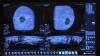
ITN Editor Dave Fornell takes a tour of some of the most innovative new trends and health information technologies (IT) on the expo floor of the Healthcare Information and Management Systems Society (HIMSS) 2016 meeting. Technologies include radiation dose management, wearables, patient engagement, admission kiosks, analytics software and imaging workflow aids.
itnTV
VIDEO: One on One with Reed A. Omary, MD, MS, Vanderbilt University Medical Center
Radiology Business | July 30, 2024
Find actionable insights to achieve sustainability and savings in radiology in this newest of ITN’s “One on One” video series with Reed A. Omary, MD, MS, Vanderbilt University Medical Center (Nashville, TN). Tune in to "Promoting the Planet's Health: Sustainability in Radiology," to hear from a recognized leader about impactful, cost-saving initiatives radiologists, associations, healthcare systems and vendors can take, and why action is imperative.
Omary, the Carol D. and Henry P. Pendergrass Professor in the VUMC Department of Radiology, is a distinguished radiologist whose commitment to driving healthcare sustainability initiatives has gained both attention and momentum. After serving as Chair of the Department of Radiology and Radiology Sciences from 2012-2023, in June, 2023, Omary stepped away from his role as Chair to pursue a sabbatical focused on climate change and sustainable healthcare. He is author of The Green Leap, a blog about making healthcare sustainable, and founder of the Greenwell Project, a sustainable healthcare non-profit. He has presented a Plenary Lecture at the Radiological Society of North America (RSNA) Annual Scientific Sessions and American College of Radiology (ACR) meetings on the topic, and continues to connect with healthcare systems, vendors and colleagues to advance the issue.
Related content:
PHILIPS MARKS MILESTONE DURING HELIUM-FREE MRI INSTALLATION IN PUERTO RICO
RSNA 2022 PLENARY SPEAKER OMARY URGES RADIOLOGISTS TO SUPPORT PATIENTS, COMMUNITIES AND THE PLANET
RSNA 2022 PANEL DISCUSSIONS FORECAST RADIOLOGY IN 2027, AND HIGHLIGHT WHY MENTORS MATTER
Conference Coverage
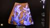
Cardiovascular Ultrasound | April 29, 2016

MD Buyline market analyst Jon Brubaker explains the new cardiac ultrasound technologies and trends he saw on the show floor at the 2016 meeting of the American College of Cardiology (ACC).
CT Angiography (CTA) | April 12, 2016

A discussion on the adoption rate of FFR-CT with Campbell Rogers, M.D., chief medical officer of HeartFlow. It is the first vendor to gain FDA approval for noninvasive, virtual fractional flow reserve measurements derived from cardiac computed tomography scans.
Related FFR-CT Content:
Clinical Applications of FFR-CT
VIDEO: Implementation and the Science Behind FFR-CT — interview with James Min, M.D.
VIDEO: Early U.S. Experience With FFR-CT in Evaluating ED Chest Pain Presentation — interview with Simon Dixon, M.D.
VIDEO: Status of FFR-CT Adoption in the United States — interview with Campbell Rogers, M.D.
Clinical Decision Support | March 24, 2016

Examples of clinical decision support software currently on the market that might be leveraged to address Stage 3 Meaningful Use from the expo floor of HIMSS 2016. Ascendian Healthcare Consulting CEO Shawn McKenzie also discusses how and why CDS should be integrated into the radiology workflow.
Patient Engagement | March 21, 2016

Examples of patient engagement technologies for medical imaging to meet health IT Stage 3 Meaningful Use requirements. Discussion includes examples from the expo floor at HIMSS 2016 and Ascendian Healthcare Consulting CEO Shawn McKenzie explaining ways radiology can leverage technology to engage patients with images, reports and radiation dose records.
PACS | March 11, 2016
Examples of technologies on the market and a discussion of what to look for in PACS and CVIS workflow efficiencies with Ascendian Healthcare Consultant Jef Williams. Editor Dave Fornell takes viewers on a tour of some of the key workflow improvements offered by health IT vendors in their software on the expo floor at Healthcare Information Management and Systems Society (HIMSS) 2016 meeting.
Related Content:
6 Key Health Information Technology Trends at HIMSS 2019
Technology Report: Enterprise Imaging
VIDEO: How to Build An Enterprise Imaging System
Enterprise Imaging 2018: Balancing Strategy and Technology
Three Resolutions Worth Keeping for a More Data-driven Radiology Practice
Proton Therapy | January 22, 2016

At ASTRO 2015, Mevion highlighted the exceptional results its customers are seeing with the Mevion S250 Series proton therapy system.
Digital Radiography (DR) | January 05, 2016

RSNA 2015 was the first time Konica Minolta and Viztek displayed as a combined company. David Widmann, president and CEO, and Joe Cermin, president of healthcare IT, explain how the merger will help clinicians and how health IT plays a major role in imaging today.
Radiation Therapy | December 21, 2015

Varian offered an overview of its new technology offerings at ASTRO 2015. These include advancements in analytics, treatment planning, adaptive therapy, clinical decision support and new tools for the integration of MRI and PET/CT into the treatment planning process.
Digital Radiography (DR) | December 14, 2015

Interview with Lori Webb, BAAS, RT, MD Buyline analyst, covering new technology introductions and trends in digital radiography (DR) systems at the Radiological Society of North America (RSNA) 2015 meeting.
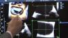
Ultrasound Imaging | December 11, 2015
Interview with Jon Brubaker, MBA, RCVT, ultrasound technology analyst, MD Buyline, explains the trends and new technology he saw at the Radiological Society of North America (RSNA) 2015 meeting.
Enterprise Imaging | December 11, 2015

Interview with Kim Garriott, principal consultant, Logicalis Inc., and Jef Williams, COO Ascendian Healthcare Consulting, explaining details of how to create an enterprise imaging system at the Radiological Society of North America (RSNA) 2015 meeting. For more information, watch the video “Six Tenets of Enterprise Imaging Strategy.”
Quality Assurance (QA) | August 20, 2015

Learn more about myQA, IBA’s unique platform that connects QA applications, people, and know-how through a central database and the Cloud. It offers full support throughout all of your QA, and enables you access to the different software modules and all of your data from one intuitive interface – anywhere and anytime.
Radiology Imaging | August 07, 2015

ITN Editor Dave Fornell shares his choices for some of the most innovative new technology on the show floor at the 2015 AHRA meeting in Las Vegas.
Radiology Imaging | August 07, 2015

Interview with Dave Fox, AHRA president, and president of the North Campus, system vice president of statewide network development, St. Vincent Health, Little Rock, Ark.
Information Technology | August 07, 2015

Interview with Melody Mulaik, MSHS, Coding Strategies Inc., Powder Springs, Ga., explains what to look for to avoid coding and reimbursement issues following the implementation of ICD-10 coding after Oct . 1, 2015.
Radiation Oncology | July 31, 2015

Joseph Deasy, Ph.D., discusses his study on dose-volume relations for late rectal bleeding in 1,001 patients from five prostate cancer cohorts with Imaging Technology News Editorial Director Melinda Taschetta-Millane.
Computed Tomography (CT) | July 30, 2015

During the American Association of Physicists in Medicine (AAPM) 57th annual meeting in Anaheim, Calif., Imaging Technology News’ editorial director Melinda Taschetta-Millane spoke with Michael F. McNitt-Gray, PhD, FAAPM, about his abstract on Size-Specific, Scanner-Independent Fetal Dose Estimates in Abdominal and Pelvic CT Examinations of Pregnant Patients, which was presented at the meeting.
Breast Imaging | July 29, 2015

During the American Association of Physicists in Medicine (AAPM) 57th annual meeting in Anaheim, Calif., graduate student researcher at the University of California/Davis Andrew M. Hernandez discussed screening and dose distribution for women with dense breasts, and its implications for mammography.
Radiation Oncology | July 28, 2015

American Association of Physicists in Medicine (AAPM) President John Boone, Ph.D., discusses the impact of imaging in medical physics, as well as key topics addressed at this year’s meeting, with Imaging Technology News Editorial Director Melinda Taschetta-Millane.
Cardiovascular Ultrasound | June 29, 2015
Interview at the American Society of Echocardiography (ASE) annual meeting with Federico Asch, M.D., M.D., FACC, FASE, associate director of the echocardiography core lab at Medstar Health Research Institute and assistant professor of medicine (cardiology) at Georgetown University.
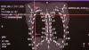
Cardiovascular Ultrasound | June 26, 2015

DAIC Editor Dave Fornell shares his choices for the most innovative new echocardiography technologies and trends at the 2015 American Society of Echocardiography (ASE) annual meeting.
Cardiovascular Ultrasound | June 26, 2015

Interview at the American Society of Echocardiography (ASE) annual meeting with Federico Asch, M.D., M.D., FACC, FASE, associate director of the echocardiography core lab at Medstar Health Research Institute and assistant professor of medicine (cardiology) at Georgetown University. He explains how ultrasound might have a roll in the future for breaking up clots and targeted delivery of gene and drug therapies.
Cardiovascular Ultrasound | June 17, 2015

Role of Interventional Echcardiography in Transcatheter Structural Heart Procedures — Rebecca Hahn, M.D., Columbia University Medical Center, is an expert in the new subspecialty of interventional echo and shares her insights at the Cardiovascular Research Foundation's (CRF) Transcatheter Valve Therapies (TVT) conference in Chicago in June.
Enterprise Imaging | June 11, 2015

With the advent of enterprise imaging, the enterprise viewer is a pivotal component that enables a single point of access to all imaging — diagnostic, clinical, mobile, EHR-driven, and medical multimedia objects - across a healthcare enterprise. Visage Imaging's General Manager, North America, Brad Levin discusses trends in the industry and the transition away from traditional PACS during SIIM 2015.
Information Technology | June 04, 2015

SIIM 2015 Program Committee Chair Richard Wiggins III, M.D., discusses social media and mHealth with ITN editorial director Melinda Taschetta-Millane, and explains what SIIM is doing to integrate it.
Information Technology | June 04, 2015

Donald Dennison, co-chair of the SIIM Hackathon committee, discusses the objectives for this event's second year at the Society for Imaging Informatics in Medicine (SIIM) 2015 annual meeting in Washington, D.C.
Information Technology | June 04, 2015

SIIM Chair David Brown discusses interoperability, and his concerns from a data security and compliance perspective, at SIIM 2015.
Information Technology | June 04, 2015

Donald Dennison, director-at-large on the Board of Directors for SIIM, shares his thoughts on "the next imaging evolution" during the Society for Imaging Informatics in Medicine (SIIM) 2015 annual meeting in Washington, D.C., and the technical and market forces that are driving this change.
Information Technology | April 23, 2015

DAIC/ITN Editor Dave Fornell shows examples of new healthcare IT technology at the 2015 HIMSS meeting that will change the future of healthcare. These include healthcare wearable devices, smart phone apps, virtual training software, population health data, and technology for patient engagement.
Information Technology | April 22, 2015

The Centers for Medicare and Medicaid (CMS) has issue a draft list Stage 3 Meaningful Use requirements. Jeff Coughlin, senior director, federal and state affairs, Health Information and Management Systems Society (HIMSS), explains what these requirements include at the HIMSS 2015 annual meeting.
April 22, 2015

During HIMSS 2015, Louis Lannum, director, ITD enterprise imaging, information technology division, Cleveland Clinic, explained in sessions how to create an enterprise imaging system that goes beyond PACS to service all imaging and data needs of departments in the hospital enterprise.
Information Technology | April 22, 2015

At HIMSS 2015, one of the biggest trends was the explosion of consumer health related wearable devices and smartphone apps and how these will integrate into the healthcare system for improved patient monitoring and patient engagement. Thomas Martin, HIMSS director of health information systems, explains this trend and where these devices will fit in during the coming years.
Enterprise Imaging | April 22, 2015

Mony Weschler, chief applications strategist and architect, application technology services, Montefiore Health System, New York, explains how he integrated enterprise imaging and mobile ECG waveform at Montefiore Health System.
Breast Density | March 27, 2015
Dense breast tissue can mask the appearance of tumors and limit the performance of mammography. When used as an adjunct to mammography, Invenia ABUS from GE Healthcare has been shown to improve invasive breast cancer detection by 55% over mammography alone.
Breast Imaging | March 24, 2015

Kristie Bobolis, M.D., program chair for the National Consortium of Breast Centers, discusses the role of genetic testing, how it is evolving, and how a physician best assess the high-risk patient.
Breast Imaging | March 24, 2015

Barbara Rabinowitz, Ph.D., MSW, RN, founder of the National Consortium of Breast Centers, talks with ITN editorial director Melinda Taschetta-Millane during NCoBC's 25th anniversary conference, and addresses the very important topic of survivorship.
Breast Density | March 24, 2015

Gary Levine, M.D., president of the National Consortium of Breast Centers, discusses breast density, updates from the legislative front and the latest breast screening technology.
Breast Imaging | March 24, 2015

Jennifer Gass, M.D., FACS, incoming president of the National Consortium of Breast Centers, discusses the impact of breast surgery on a woman's sense of sexuality, and her future goals during her presidency of NCBC.

© Copyright Wainscot Media. All Rights Reserved.
Subscribe Now