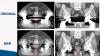
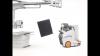
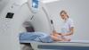
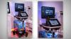
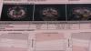
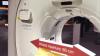
VIVIX-S 1417N is a multi-purpose portable flat panel detector for addressing busy workflow in medical facilities. Get more information on our advanced detectors and software lineup at www.vieworks.com.
Watch the VIDEO here.
Find actionable insights to achieve sustainability and savings in radiology in this newest of ITN’s “One on One” video series with Reed A. Omary, MD, MS, Vanderbilt University Medical Center (Nashville, TN). Tune in to "Promoting the Planet's Health: Sustainability in Radiology," to hear from a recognized leader about impactful, cost-saving initiatives radiologists, associations, healthcare systems and vendors can take, and why action is imperative.
Omary, the Carol D. and Henry P. Pendergrass Professor in the VUMC Department of Radiology, is a distinguished radiologist whose commitment to driving healthcare sustainability initiatives has gained both attention and momentum. After serving as Chair of the Department of Radiology and Radiology Sciences from 2012-2023, in June, 2023, Omary stepped away from his role as Chair to pursue a sabbatical focused on climate change and sustainable healthcare. He is author of The Green Leap, a blog about making healthcare sustainable, and founder of the Greenwell Project, a sustainable healthcare non-profit. He has presented a Plenary Lecture at the Radiological Society of North America (RSNA) Annual Scientific Sessions and American College of Radiology (ACR) meetings on the topic, and continues to connect with healthcare systems, vendors and colleagues to advance the issue.
PHILIPS MARKS MILESTONE DURING HELIUM-FREE MRI INSTALLATION IN PUERTO RICO
RSNA 2022 PLENARY SPEAKER OMARY URGES RADIOLOGISTS TO SUPPORT PATIENTS, COMMUNITIES AND THE PLANET
RSNA 2022 PANEL DISCUSSIONS FORECAST RADIOLOGY IN 2027, AND HIGHLIGHT WHY MENTORS MATTER
VIVIX-S 1417N is a multi-purpose portable flat panel detector for addressing busy workflow in medical facilities. Get more information on our advanced detectors and software lineup at www.vieworks.com.
Watch the VIDEO here.
Scott Schwartz, M.D., interventional radiologist and program director for IR residencies and the vascular and interventional radiology fellowship at Henry Ford Hospital, explains how the department uses Yttrium-90 (Y90) embolization therapy to treat liver cancer.
Find more content on Henry Ford Hospital
Related Articles on Y-90 Radiotherapy:
Current Advances in Targeted Radionuclide Therapy
A Look Ahead in Targeted Radionuclide Therapy
Radioactive Bead Therapy Now Used for Head, Neck Tumors
NCCN Guidelines Recommend Y-90 Microspheres for Metastatic Colorectal Cancer Treatment
Benjamin Movsas, M.D., chair of radiation oncology at Henry Ford Cancer Institute, describes the benefits the department has realized from using the first FDA-approved magnetic resonance imaging (MRI)-guided radiation therapy system to allow real-time treatment tracking.
Watch the related VIDEO: MRI-Guided Radiation Therapy Trial for Pancreatic Cancer.
Register to view a webinar on the ViewRay MRI-guided radiation therapy system.
Find more content on Henry Ford Hospital
Carri Glide-Hurst, Ph.D., director of translational research, radiation oncology at Henry Ford Health System, describes how the department uses magnetic resonance imaging (MRI) scans to create synthetic computed tomography (CT) images for use in radiation therapy treatment planning.
Also watch the VIDEOs Using Advanced CT to Enhance Radiation Therapy Planning and MRI-Guided Radiation Therapy Trial for Pancreatic Cancer, which also feature Glide-Hurst.
Carri Glide-Hurst, Ph.D., director of translational research, radiation oncology at Henry Ford Health System, describes how the department uses advanced computed tomography (CT) software and techniques to improve radiation therapy treatment plans.
Related Content With Glide-Hurst:
WEBINAR: Advances in CT to Enhance Radiation Therapy Planning
VIDEO: Clinical Use of the ViewRay MRIdian Linac System at Henry Ford
VIDEO: Deriving Synthetic CT Images for Radiotherapy Treatment Planning
VIDEO: MRI-Guided Radiation Therapy Trial for Pancreatic Cancer
Carri Glide-Hurst, Ph.D., and Benjamin Movsas, M.D., discuss Henry Ford Hospital's involvement in a national clinical trial assessing the effectiveness of magnetic resonance imaging (MRI)-guided radiation therapy in pancreatic cancer.
Watch the related VIDEO: Clinical Use of the ViewRay MRIdian Linac System at Henry Ford — Interview with Benjamin Movsas, M.D.
Find more content on Henry Ford Hospital
Register to view a webinar on the ViewRay MRI-guided radiation therapy system.
A discussion with Ehsan Samei, Ph.D., DABR, FAAPM, FSPIE, director of the Duke University Clinical Imaging Physics Group and head of the Duke medical physics graduate program. He spoke on this topic at the American Association of Physicists in Medicine (AAPM) 2018 meeting.
Haojie Wang, M.D., director of advanced cardiovascular MRI and a member of the heart valve clinic at Baylor Scott White Heart Hospital, Dallas, explains the utility of magnetic resonance imaging (MRI) in cardiology and the implementation of the cardiac MRI program at his hospital.
A discussion with Martin Vallieres, Ph.D., post-doctoral fellow at McGill University, Montreal, Canada. He spoke on this topic in sessions at the American Association of Physicists in Medicine (AAPM) 2018 meeting. He explains radiomics and how it can be used in both medical imaging and radiation therapy.
Read the related article "Hidden Information Behind Imaging Tests for Cancer May Unlock New Approaches to Radiation Therapy."
Read the related article "Computer Program Beats Physicians at Brain Cancer Diagnoses."
Lawrence Tanenbaum, M.D., FACR, vice president and director of advanced imaging at RadNet, discusses the latest research on the effects of gadolinium contrast retention following magnetic resonance imaging (MRI) exams, and how facilities can best manage its use.
Sheila Sferrella, president of Regents Health Resources and Bill Finerfrock, president of Capitol Associates, discuss the national progress in transitioning from computed radiography (CR) to digital radiography (DR) systems. Sferrella is the chair and Finerfrock is a member of the AHRA Regulatory Affairs Committee.
Watch the related Technology Report on Digital Radiography Systems
Watch part 1 of the conversation with Sferrella and Finerfrock on clinical decision support updates
Ehsan Samei, Ph.D., DABR, FAAPM, FSPIE, director of the Duke University Clinical Imaging Physics Group and head of the Duke medical physics graduate program, explained this new program at the American Association of Physicists in Medicine (AAPM) 2018 meeting.
Robert Junk and Tobias Gilk, MRSO, MRSE, of architectural firm RAD-Planning, discuss the different types of safety hazards associated with magnetic resonance imaging (MRI) and how to assess your own protocols to find and eliminate inefficiencies that could lead to safety hazards.
Watch the related VIDEO New App Improves MRI Safety For Implantable Devices
Read the related article "Closing the Loopholes in MRI Safety"
A discussion with Steve Jiang, Ph.D., director of the medical artificial intelligence and automation lab and vice-chair of the Department of Radiation Oncology, University of Texas Southwestern. He spoke in sessions at AAPM 2018 about how AI will change treatment planning, radiation oncology and medical imaging.
Watch the VIDEO: Real-world Implementation of Deep Learning for Treatment Planning — a discussion with Kevin Moore, Ph.D., DABR, deputy director of medical physics and associate professor, University of California San Diego, about his daily clinical use of an artificial intelligence treatment planning software.
Other coverage from the AAPM 2018 meeting.
#AAPM2018
A discussion with Patricia Dickson, LRT(CT), director of imaging and outpatient services, Capital Cardiology Associates, Albany, N.Y., and Nikki Weber, a lead CT technologist at the Mayo Clinic, Rochester, Minn. They presented in sessions at the Society of Cardiovascular Computed Tomography (SCCT) 2018 meeting.
Watch the related VIDEO: How to Prepare a Patient for a Cardiac CT Scan
ITN Associate Editor Jeff Zagoudis speaks with Karen Hou, M.D., breast radiologist at Northwestern Medicine Central DuPage Hospital in Winfield, Ill., about the hospital's use of new technology and innovative practices. Read more about the clinic the article "High-Risk Clinic Arms Patients Against Breast Cancer."
See more content from ITN's visit to Northwestern Medicine Central DuPage Hospital.
Related Breast Imaging Content:
VIDEO: The Impact of Breast Density Technology and Legislation
High-Risk Clinic Arms Patients Against Breast Cancer
Greater Evidence, Payment Expansion Driving Tomosynthesis Adoption
Technology to Watch in Breast Imaging
VIDEO: Developments in Breast Density Legislation and Screening Guidelines
The Transition to 3-D Breast Imaging
ITN Associate Editor Jeff Zagoudis explores how the mobile stroke unit (MSU) program at Northwestern Medicine Central DuPage Hospital in Winfield, Ill., is changing the paradigm of care for stroke patients in the western suburbs of Chicago and beyond.
Take a look around with a 360 degree view inside the mobile stroke unit ambulance.
ITN Editorial Director Melinda Taschetta-Millane speaks to DenseBreast-info Executive Director JoAnn Pushkin as the website celebrates three years as an information resource on breast density for patients and providers. Pushkin also discusses the latest developments in breast density notification and screening guidelines.
Related Dense Breast Imaging Content:
VIDEO: The Impact of Breast Density Technology and Legislation
High-Risk Clinic Arms Patients Against Breast Cancer
Greater Evidence, Payment Expansion Driving Tomosynthesis Adoption
Technology to Watch in Breast Imaging
The Transition to 3-D Breast Imaging
Fujifilm details its latest innovations in women's health including its ASPIRE Cristalle mammography system with digital breast tomosynthesis software option and its Synapse 5 PACS and VNA, enabling secure, easy-to-manage storage and access to the complete patient imaging record throughout the healthcare enterprise.
FUJIFILM Medical Systems U.S.A., Inc. and FUJIFILM SonoSite Inc., are offering a full-suite pediatric solutions portfolio, complete with digital radiography (DR), healthcare IT and point-of-care ultrasound to serve the world's most unique patients.
David Widmann, president and CEO of Konica Minolta, looks at what the future of healthcare can bring to its customers, focusing on the imaging space.
Kiyotaka Fujii, global healthcare senior executive officer and president of Konica Minolta, discusses the company's medical strategy as it grows into precision medicine.
ITN Associate Editor Jeff Zagoudis demonstrates several mobile apps designed to help pediatric patients learn what an MRI exam is like to help calm their fears and make it easier to conduct the exam. Read the article "Pediatric MRI Calming Techniques"
ITN Editor Dave Fornell previews the launch of augmented reality (AR) technology in the January/February 2018 issue of ITN. Augmented reality brings new depth to print content through your smartphone by connecting to related videos and other resources.
Mindray recently featured a new upgrade for its premium Resona 7 ultrasound system at the Radiological Society of North America annual conference. The new upgrade is based on the system’s core ZONE Sonography Technology (ZST+), which enables software upgrades for new applications such as contrast enhanced ultrasound (CEUS), Sound Touch shear wave elastography and 10 new transducer options. Other imaging, software, and calculation enhancements include fetal CNS “Smart Planes,” “Smart Pelvis” and a dedicated pelvic congestion syndrome package. The new transducers include C6-2GU interventional/intraoperative; C11-3U microconvex; P10-4U and P7-3U phased pediatric cardiac; P7-3TU adult transesophageal; L16-4HU; SC6-1U and DE10-3WU (3D). There are also two new CW transducers, CW2S and CW5S, for the support of shared service. The new upgrade is expected to be available to Resona 7 customers early next year. For more information: www.mindraynorthamerica.com
ITN and DAIC Editor Dave Fornell shows several examples of how vendors are incorporating AI, deep learning algorithms into their medical imaging information system and the modalities themselves to speed workflow, improve imaging accuracy, improve reimbursements, monitor analytics in real time and eliminate the need for humans to do tedious, time consuming tasks. These examples were seen on the show floor at at the Radiological Society of North America (RSNA) 2017 meeting. For more on deep learning, watch the VIDEO "Deep Learning is Key Technology Trend at RSNA 2017," and an interview with Adam Flanders, M.D., chair of the RSNA Radiology Informatics Committee, in the VIDEO "How Utilization of Artificial Intelligence Will Impact Radiology." ITN also created an indepth VIDEO Technology Report — Artificial Intelligence at RSNA 2017 with interviews with numerous AI vendors.
Watch the VIDEO “Editor’s Choice of the Most Innovative New Imaging Technology at RSNA 2017.”
Max Wintermark, M.D., professor of radiology and chief of neuroradiology, Stanford Hospital and Clinics, discussed MRI neurology quantification apps at the Radiological Society of North America (RSNA) 2017 meeting. Read the article “Software Advances in MRI Technology.” Watch the VIDEO “Neuroimaging Advances.”
ITN and DAIC Editor Dave Fornell takes a tour of some of the most interesting new medical imaging technologies on the expo floor at the 2017 Radiological Society of North America (RSNA) meeting. Watch the editor’s choice video for new health IT technologies at HIMSS 2017, new advances in cardiac ultrasound at ASE 2017, and CT advances at the SCCT 2017 meeting.
Nancy M. Cappello, Ph.D., executive director and founder, Are You Dense Inc. and Are You Dense Advocacy. Read the article “The Evolution of Breast Imaging Technology.” Watch the VIDEO “Implementing Advanced Breast Imaging Technology.”
A post-game roundup by ITN Contributing Editor Greg Freiherr and ITN Editor Dave Fornell on the key trend of artificial intelligence seen on the show floor at the Radiological Society of North America (RSNA) 2017 meeting.
For more on artificial intelligence at RSNA 2017, watch the VIDEO "How Utilization of Artificial Intelligence Will Impact Radiology." For more trends in imaging, watch the VIDEO "Key Imaging Technology Trends at RSNA 2017." ITN also created an indepth VIDEO Technology Report — Artificial Intelligence at RSNA 2017 with interviews with numerous AI vendors.
Emanuel Kanal, M.D., director of MRI services and professor of radiology and neuroradiology at the University of Pittsburgh Medical Center, discusses the heightened concerns about gadolinium, which may cause adverse effects in some patients. Watch the VIDEO: How Serious is MRI Gadolinium Retention in the Brain, Body? with Max Wintermark, M.D. Read the articles “FDA: No Harm in MRI Gadolinium Retention in the Brain,” and “European Medicines Agency Issues Update on Gadolinium Contrast Agents.”
A post-game roundup by ITN Contributing Editor Greg Freiherr and ITN Editor Dave Fornell on the trends and new tech seen on the show floor at the Radiological Society of North America (RSNA) 2017 meeting. The biggest overarching trend for this year at RSNA was the explosion of artificial intelligence integration into imaging systems and medical imaging information technology. For more on the AI trend, watch the VIDEO "How Utilization of Artificial Intelligence Will Impact Radiology," with Adam Flanders, M.D.
Emanuel Kanal, M.D., director of MRI services and professor of radiology and neuroradiology at the University of Pittsburgh Medical Center, explains the new mobile application he developed, which lets users visually model the forces at work during an MRI exam on patients with implanted medical devices, at the 2017 Radiological Society of North America (RSNA) meeting.
Read the related article “The Changing Relationship Between MRIs and Pacemakers.”
Max Wintermark, M.D., professor of radiology and chief of neuroradiology, Stanford Hospital and Clinics, discussed MRI contrast safety issues at RSNA 2017. Read the article “FDA: No Harm in MRI Gadolinium Retention in the Brain,” and “European Medicines Agency Issues Update on Gadolinium Contrast Agents.” Watch the VIDEO “Big Concerns Remain for MRI Gadolinium Contrast Safety at RSNA 2017,” and interview with Emanuel Kanal, M.D.
Dianna Bardo M.D., director of body MR and co-director of the 3-D Innovation Lab at Phoenix Children's Hospital, discusses how 3-D printing and other advanced imaging modalities can help improve outcomes in complex cases. Read the article “The Use of 3-D Printing in Cardiology.” Watch the WEBINAR “Innovation and Success in 3D-inspired Development of the Business and Clinical Practice,” presented by Bardo.
Martin Yaffe, Ph.D., FAAPM, senior scientist, physical sciences/imaging research, Sunnybrook Health Sciences Centre, and professor of medical biophysics and medical imaging, University of Toronto, discusses how contrast-enhanced mammography can improve breast cancer detection at the 2017 Radiological Society of North America (RSNA) meeting.
Watch the VIDEO “What is New in Breast Imaging Technology,” with Stamatia Destounis, M.D.
Watch the VIDEO "Implementing Advanced Breast Imaging Technology," with Gerry Kolb, JD.
Stamatia Destounis, M.D., FACR, associate professor, University of Rochester School of Medicine, attending radiologist, Elizabeth Wende Breast Care, explains recent advances in mammography and dense breast imaging at the 2017 Radiological Society of North America (RSNA) meeting.
The Evolution of Breast Imaging Technology.
VIDEO: Implementing Advanced Breast Imaging Technology.
VIDEO: Use of Breast MRI Screening in Women With Dense Breasts
VIDEO: Explaining Dense Breasts — Interview with Christiane Kuhl, M.D.
Improving Clinical Image Quality for Breast Imaging
Breast Imaging in the Age of Coronavirus
VIDEO: Use of Breast MRI Improved Cancer Detection in Dense Breasts in Dutch Study — Interview with Gillian Newstead, M.D.
Dee Dee Wang, M.D., Director, Structural Heart Imaging at Henry Ford Hospital, Detroit, explains how her center uses 3-D printing and computer aided design (CAD) software to improve patient outcomes. She spoke to ITN at the 2017 Transcatheter Cardiovascular Therapeutics (TCT) annual meeting. Read the articles “The Use of 3-D Printing in Cardiology” and “Henry Ford Hospital Study Shows 3-D Imaging Improves Fixing Broken Hearts.”
The new Visicoil MR is a helically-wound, flexible linear fiducial marker. The Visicoil MR hollow core design and multiple sizes mitigates the CT artifact often created by traditional gold seeds, offering you clearer visualization of the tissue to aid in faster and more accurate contouring. Visicoil MR's linear flexible design fixates and remains stable in soft tissue, giving you the confidence in accurate marker localization. Visicoil MR is available in some of the smallest needle sizes available for fiducial markers, including 21g and 22g needles. The benefits of smaller needles are improved patient safety and comfort during implantation. The smaller needles may also allow for faster CT sim after implant and ultimately treatment for your patients. The new Visicoil MR with enhanced MRI visibility allows you to see the marker more easily in MRI, both in T1 and T2. This makes for easier and faster MR/CT fusion.
Prem Soman, M.D., director of nuclear cardiology at the Heart and Vascular Institute, University of Pittsburgh, and president-elect of the American Society of Nuclear Cardiology (ASNC), explained advances in PET and SPECT imaging and the learning curve involved in reading scans from the new CZT SPECT cameras. Watch the VIDEO: Trends in Nuclear Cardiology Imaging, an iknterview with David Wolinsky, M.D., director of nuclear cardiology at Cleveland Clinic Florida. Read the related article "Advances in Cardiac Nuclear Imaging."